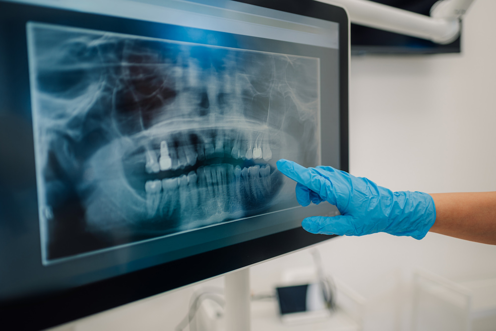
A missing tooth doesn’t create the same treatment problem everywhere in the mouth. Front teeth shape your smile and help guide certain bite movements, while back teeth do much of the heavy chewing. Anyone researching dentist implants in Murrieta should know the location of the gap is one of the first details that influences how the replacement is planned.
The site affects the pressure the new tooth will handle, the amount of bone available, the surrounding gum tissue, and the nearby anatomy that needs to be protected. Looking closely at the exact area shows the surgeon how the site shapes the dental implant plan.
Are front and back dental implants planned differently?
Yes. The part of the mouth being restored changes what the replacement has to do. Front teeth are highly visible and shape the appearance of your smile, while back teeth take on more of the pressure created during chewing. The plan has to reflect those differences.
Front teeth require a precise fit
In the front of the mouth, position matters for more than function. Even a small shift can affect how the crown lines up with the neighboring teeth or how naturally it meets the gumline. The bone along the outer surface of the jaw can also be thinner in this area, leaving less room to work with.
The fixture therefore has to sit within the available bone while giving the future crown enough space to look natural. Gum shape matters too because the tissue around a front tooth is visible when you smile.
The plan changes when the gap includes more than one tooth. Replacing several teeth requires a different approach because the surgeon has to account for how multiple replacements will work together across a larger area.
Back teeth have to handle more pressure
Molars and premolars do much of the heavy chewing, so their replacements need to be planned with that workload in mind. While bone availability still matters, the surrounding anatomy can place additional limits on the space available.
In the upper jaw, the back teeth sit close to the maxillary sinus. In the lower jaw, the nearby nerve can affect how much room there is for placement. The surgeon uses those boundaries to decide what size fixture will fit safely and whether the site needs any preparation beforehand.
When many teeth are missing, full-arch treatment can use different fixture counts. All-on-4, All-on-6, and other All-on-X approaches use different layouts based on the anatomy and the prosthesis being supported.
Why does the amount of chewing force matter?
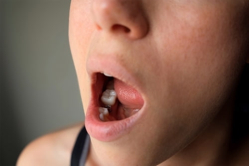 A replacement tooth has to function as part of the bite and fill the empty space. Back teeth take greater vertical pressure because they grind food. Front teeth experience more directional forces during biting and jaw movement.
The surgeon considers how that pressure will travel through the crown and into the jaw. Available bone, replacement size, bite relationship, and jaw movement all help shape the plan.
Several details influence the amount of force a site receives:
- Tooth location: Molars handle heavier chewing than incisors.
- Bite pattern: The way the upper and lower teeth meet changes where pressure is concentrated.
- Clenching or grinding: Repeated heavy loading can place more stress on the restoration and supporting bone.
- Number of missing teeth: A longer bridge or full-arch prosthesis distributes force differently from a single crown.
Dental implants are designed to restore the ability to chew, so bite function is considered from the beginning. The position has to work for the way that part of the mouth is used every day. The way those forces are handled also comes down to precise positioning. Placement depends on angle and depth, along with how much bone is available in that particular area.
Does bone anatomy vary depending on where the missing tooth is located?
Yes, because the jaw isn’t uniform from front to back. Bone thickness and density change by location, and nearby anatomy can create different limits from one site to another. Two missing teeth in the same patient can therefore require very different plans.
The main differences involve:
- Upper front jaw: The outer bone can be thin, and the visible gumline makes contour especially important.
- Upper back jaw: The sinus can reduce the amount of vertical bone available above the missing tooth.
- Lower front jaw: The ridge is often narrower, so width becomes an important part of planning.
- Lower back jaw: The inferior alveolar nerve can limit how far a fixture can extend into the jaw.
Three-dimensional imaging helps the surgeon measure these areas before surgery and see how much room is available around the planned site.
Bone also changes after a tooth is removed
The socket doesn’t stay exactly the same after an extraction. The ridge remodels as it heals, and some of its width and height can decrease over time. Research has found that post-extraction bone changes are often greatest early in healing, although the amount varies by site and patient.
People searching how to encourage bone growth after tooth extraction are often trying to understand how enough bone can be preserved for a future replacement. Depending on the site, the surgeon may discuss socket preservation or bone grafting to help maintain or rebuild the ridge.
The long-term side effects of tooth extraction can include continued changes in the ridge and less bone available at the missing-tooth site. Treatment can still be possible later. The current anatomy needs to be evaluated before a plan is made.
An older extraction site can still be evaluated
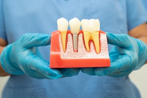 Patients sometimes wonder when is it too late to get dental implants after living with a gap for years. There’s no single cutoff based on time alone. The main question is whether enough healthy bone remains and whether the site can be prepared safely if extra support is needed.
One healed area may lose more width or height than another. Imaging shows what is available and whether grafting or a different fixture size should be considered before treatment moves forward.
Who can I speak with about dentist implants in Murrieta?
A consultation gives you a clearer picture of what the missing-tooth site allows and what type of replacement makes sense for that part of your mouth. Temecula Center for Dental Implants provides individualized planning with Dr. Dmitry Tsvetov, a board-certified oral and maxillofacial surgeon experienced in both single-tooth and full-arch treatment.
For patients across Murrieta, from neighborhoods near Murrieta Creek to other parts of the city, advanced imaging helps show how much bone is available and what nearby anatomy needs to be considered.
Contact the office today to schedule a consultation and get a treatment plan built around your specific tooth location.